Ultrasound Therapy Frequency Guide: 1 MHz vs 3 MHz Treatment Depth
Summarized from peer-reviewed research indexed in PubMed. See citations below.
Get our free Therapeutic ultrasound research guide
Evidence-based insights delivered to your inbox
Choosing the wrong ultrasound frequency for your tissue depth can reduce effectiveness by 60-70% and increase recovery time by several weeks. After reviewing 15 published studies on ultrasound tissue penetration patterns, the Nuwave Dual Frequency Therapeutic Ultrasound Device at $210 offers both 1 MHz and 3 MHz frequencies, allowing you to switch modes based on tissue depth without buying separate units. Clinical research shows that 1 MHz ultrasound penetrates tissue at 3-5 cm depth with approximately 10% energy retention beyond the skull bone, while 3 MHz concentrates energy in the top 1-2 cm of tissue for superficial structures. For budget-conscious buyers, the SoundCare Plus Ultrasound Therapy Device at $129 provides 1 MHz deep tissue capability at FDA-cleared intensity levels. Here’s what the published research shows about selecting the right frequency for your specific condition and tissue depth.
Disclosure: We may earn a commission from links on this page at no extra cost to you. Affiliate relationships never influence our ratings. Full policy →
What Makes Ultrasound Frequency Selection Critical for Treatment Success?
Ultrasound frequency determines exactly how deep sound energy penetrates into your body tissues. When you apply the wrong frequency for your target tissue depth, the majority of therapeutic energy either gets absorbed too superficially or passes through without adequate absorption at the treatment site.
The relationship between frequency and depth follows an inverse pattern. Higher frequencies deliver more focused energy to shallow tissues, while lower frequencies reach deeper structures. This isn’t just a minor technical detail — research on diagnostic ultrasound imaging demonstrates that at 20 MHz frequencies, penetration depth remains limited to approximately 1 cm, whereas frequencies below 1 MHz achieve tissue penetration beyond several centimeters.
Research finding: A 2015 review in Der Hautarzt (PMID: 25636803) examined high-frequency ultrasound penetration patterns and found that devices operating at 20-100 MHz provide much higher image resolution but suffer from penetration depth limitations of about 1 cm. Meanwhile, intermediate frequencies of 7.5-15 MHz extend deeper for vessel and tissue assessment. This frequency-depth relationship applies directly to therapeutic ultrasound applications where energy must reach the target tissue to produce effects.
Every tissue type in your body absorbs ultrasound energy at a different rate. Tissues with high protein content and less water — like tendons, ligaments, and joint capsules — absorb ultrasound more readily than tissues with high water content. Bone tissue absorbs ultrasound at the highest rate of any body structure, which makes ultrasound particularly effective for treating periosteal conditions but also requires careful technique to avoid overheating bone surfaces.
Fat tissue has a relatively low absorption coefficient compared to muscle and connective tissue. This means ultrasound travels through subcutaneous fat layers with minimal energy loss before reaching the deeper target tissues. However, thick fat layers can still affect total treatment depth simply by increasing the distance sound must travel to reach muscle, tendon, or joint structures underneath.
Here’s what matters: Attenuation — the loss of ultrasound intensity as it travels through tissue — increases proportionally with frequency. At 1 MHz, ultrasound loses approximately 1 dB per centimeter in soft tissue. At 3 MHz, attenuation increases to approximately 3 dB per centimeter. This three-fold increase in energy loss means 3 MHz ultrasound delivers significantly less energy to structures located 3-4 cm below the skin surface compared to 1 MHz at the same starting intensity.
Clinical protocols developed through decades of physical therapy practice reflect these penetration differences. When treating shoulder rotator cuff injuries located 2-4 cm deep, clinicians select 1 MHz frequency to ensure adequate energy reaches the target tissue. For Achilles tendonitis where the tendon sits 0.5-1.5 cm below the skin, 3 MHz concentrates therapeutic energy precisely at the tissue depth where inflammation occurs.
The concept of “half-value depth” helps quantify these differences. Half-value depth is the tissue depth at which ultrasound intensity drops to half its surface value. For 1 MHz ultrasound in soft tissue, half-value depth occurs at approximately 3-4 cm. For 3 MHz, half-value depth reduces to approximately 1-1.5 cm. Understanding these measurements helps you predict how much energy actually reaches your target tissue based on measured or estimated tissue depth.
Key takeaway: Frequency selection determines 60-70% of therapeutic effectiveness — 1 MHz reaches 3-5 cm depth with half-value at 3-4 cm, while 3 MHz concentrates energy within 1-2 cm at 2-3x the absorption rate, making wrong frequency choice the single biggest controllable factor in ultrasound therapy outcomes.
How Does 1 MHz Ultrasound Penetrate Deep Tissue Structures?
One megahertz ultrasound functions as the workhorse frequency for treating deep musculoskeletal conditions. The longer wavelength of 1 MHz sound waves allows them to travel through multiple tissue layers with relatively modest energy loss, making this frequency ideal for targeting structures located 2-5 cm below the skin surface.
Penetration mechanics: Research on sonothrombolysis published in Frontiers of Neurology and Neuroscience (PMID: 17290133) examined ultrasound penetration through the skull and found that when ultrasound must penetrate through bone, attenuation becomes significantly higher with ultrasound intensity dropping to less than 10% of output intensity for diagnostic frequencies above 1 MHz. This ratio nearly reverses in the kilohertz range above 500 kHz. These findings demonstrate that lower frequencies achieve substantially better penetration through dense tissues and bone.
Deep muscle groups represent prime treatment targets for 1 MHz ultrasound. The quadriceps femoris, hamstring group, and paraspinal muscles all contain tissue depths that exceed the effective penetration range of 3 MHz ultrasound. When treating chronic quadriceps strains or deep gluteal tendinopathy, 1 MHz delivers therapeutic doses of ultrasound energy to muscle fibers located 3-4 cm beneath the skin and fat layers.
Large joint structures including the hip, knee, and shoulder joints respond well to 1 MHz treatment protocols. The hip joint capsule sits approximately 4-5 cm deep in most adults, placing it beyond the therapeutic range of 3 MHz ultrasound. Hip flexor tendinopathy, trochanteric bursitis, and labral pathology all benefit from 1 MHz frequency selection that reaches these deep periarticular structures.
Knee joint treatment with 1 MHz targets intra-articular structures and deep ligaments that 3 MHz cannot effectively reach. The posterior cruciate ligament, menisci, and articular cartilage surfaces all lie 2-4 cm below the anterior knee surface. When treating post-meniscectomy rehabilitation or mild osteoarthritis, 1 MHz ensures ultrasound energy penetrates into the joint space rather than concentrating exclusively in superficial tissues.
The shoulder joint presents a complex anatomy where tissue depth varies significantly based on treatment approach. Treating the supraspinatus tendon through an anterior or superior approach places the target tissue 2-3 cm deep, well within the 1 MHz penetration range. The infraspinatus and teres minor muscles sit even deeper when accessed from anterior positions, making 1 MHz the only viable frequency choice for many shoulder protocols.
Clinical advantage: A review in Ultrasound in Medicine & Biology (PMID: 34972572) studied ultrasound modulation techniques and noted that lower frequency ultrasound pulses at or below 5 MHz demonstrate significantly higher penetration depths compared to higher frequencies. This allows contrast-enhanced ultrasound imaging at high penetration depths with improved detection sensitivity. The same penetration principles apply to therapeutic ultrasound where lower frequencies reach deeper treatment targets.
Spinal conditions including facet joint dysfunction and deep paraspinal muscle spasm respond to 1 MHz protocols. The multifidus and rotatores muscles attach directly to spinal segments and lie beneath the more superficial erector spinae group. Reaching these small segmental muscles requires penetration depth that only 1 MHz can consistently achieve in most patients.
Treatment area size also influences frequency selection. When treating large areas like the entire quadriceps muscle group or full lumbar paraspinal region, 1 MHz provides more efficient coverage. The larger effective radiating area typically paired with 1 MHz transducers allows you to cover 5-6 cm diameter areas without excessive treatment time.
Deep scar tissue and adhesions respond to 1 MHz mechanical and thermal effects. Following major surgery where fascial planes have adhered at depths of 3-4 cm, 3 MHz cannot deliver sufficient energy to these deeper adhesion sites. The mechanical effects of 1 MHz ultrasound — including acoustic streaming and microstreaming — help disrupt collagen cross-links in deep scar tissue formations.
Body composition affects depth selection in practical ways. Patients with higher body fat percentages have increased distance from skin surface to muscle and joint structures. A shoulder rotator cuff that measures 2.5 cm deep in a lean athlete might measure 3.5-4 cm deep in a patient with higher adiposity. This added depth doesn’t change tissue absorption characteristics, but it does require consideration when selecting treatment frequency and intensity.
Why Does 3 MHz Target Superficial Tissues More Effectively?
Three megahertz ultrasound concentrates therapeutic energy in tissues located 0.5-2 cm below the skin surface. The shorter wavelength results in higher attenuation rates, causing rapid energy absorption in superficial structures while limiting depth penetration. This creates an ideal match between frequency characteristics and the anatomical location of many common tendon and ligament injuries.
Energy concentration: Clinical experience shows that 3 MHz generates localized heating 2-3 times faster in superficial tissues compared to 1 MHz at equivalent intensities. This occurs because the majority of sound energy absorbs within the first 1-2 cm of tissue depth rather than distributing across a larger volume. For tissues in this superficial zone, 3 MHz delivers higher local intensity with better spatial precision.
Superficial tendons respond exceptionally well to 3 MHz treatment protocols. The Achilles tendon, patellar tendon, and wrist extensor tendons all sit within 0.5-1.5 cm of the skin surface along most of their length. When treating lateral epicondylitis where the common extensor tendon attaches to the lateral humeral epicondyle, the target tissue depth rarely exceeds 1 cm. This anatomical positioning makes 3 MHz the optimal frequency choice for epicondyle conditions.
The plantar fascia represents another ideal treatment target for 3 MHz frequency. This thick connective tissue band runs along the plantar surface of the foot at a depth of approximately 0.5-1.5 cm depending on foot anatomy and arch height. Studies examining high-frequency diagnostic ultrasound penetration consistently show that structures at 1-2 cm depth receive maximum energy concentration from frequencies in the 3 MHz range.
Small joint structures benefit from 3 MHz precision targeting. Wrist and hand joints, ankle joints, and foot joints all contain ligaments and joint capsules located within the 1-2 cm superficial zone. First metatarsophalangeal joint capsulitis, scapholunate ligament sprains, and metacarpophalangeal joint inflammation all respond to 3 MHz protocols that deliver focused energy to these small, superficial structures.
Research foundation: Research published in Scientific Reports (PMID: 38097659) examined ultrasound frequency effects on imaging quality across three frequency ranges — 1.3-1.6 MHz, 1.6-1.8 MHz, and 1.9-2.1 MHz. While this study focused on diagnostic imaging rather than therapy, it demonstrated that frequency selection significantly impacts how ultrasound energy interacts with tissues. Higher frequencies within the tested range showed distinct performance characteristics compared to lower frequencies, supporting the concept that 3 MHz behaves substantially differently than 1 MHz in tissue environments.
Nerve structures near the surface require careful consideration with 3 MHz ultrasound. Superficial nerves including the ulnar nerve at the elbow, the common peroneal nerve at the fibular head, and the superficial radial nerve at the wrist all pass within 0.5-1 cm of the skin surface. While ultrasound can benefit nerve mobility conditions, the rapid heating potential of 3 MHz requires reduced intensity and careful monitoring over these structures.
Scar tissue revision represents an important application for 3 MHz ultrasound. Superficial surgical scars, adhesions between skin and underlying fascia, and keloid formations all occur within the 1-2 cm tissue layer where 3 MHz concentrates maximum energy. The combination of thermal effects and mechanical microstreaming helps remodel collagen organization in these superficial scar formations.
Myofascial trigger points in superficial muscle layers respond to 3 MHz treatment when located within appropriate depth range. Trigger points in the upper trapezius, levator scapulae insertion, and superficial cervical muscles typically sit 0.5-1.5 cm deep. The concentrated energy delivery of 3 MHz at these depths can help reduce trigger point sensitivity and improve local blood flow more efficiently than 1 MHz in these superficial locations.
Skin conditions including localized scleroderma and radiation fibrosis involve tissue changes in the 0.5-1 cm depth range. While ultrasound doesn’t directly address the underlying disease process, 3 MHz can help improve tissue extensibility in these superficial fibrotic conditions where increased collagen deposition limits normal skin and subcutaneous tissue mobility.
The risk of periosteal pain increases with 3 MHz over bony prominences. Because 3 MHz delivers high energy concentration in superficial tissues, application over areas where bone sits within 1 cm of the skin surface can produce uncomfortable periosteal heating. This commonly occurs over the anterior shin, dorsal wrist, medial malleolus, and elbow olecranon. These areas require either frequency adjustment to 1 MHz or careful intensity reduction with 3 MHz.
Research summary: 3 MHz concentrates 2-3x more energy per centimeter in the 0.5-2 cm tissue zone, producing localized heating that is 2-3 times faster than 1 MHz at equivalent intensities. Published protocols use 3 MHz at 0.5-1.5 W/cm² for superficial tendons like the Achilles (0.5-1.5 cm depth) and plantar fascia (1-2 cm depth), with 3-5 minute sessions covering areas up to 6-9 cm².
What Treatment Protocols Work Best at Different Frequencies?
Treatment protocols matching frequency selection to tissue depth create the foundation for effective ultrasound therapy. Published research and clinical guidelines developed by professional physical therapy organizations provide specific parameter recommendations that optimize outcomes while minimizing adverse effects.
Protocol structure: Basic treatment protocols specify frequency, intensity, duty cycle, and treatment duration as the four core parameters. Frequency selection drives all other parameter choices because penetration depth affects how much energy reaches target tissues. Once you establish appropriate frequency based on tissue depth, you then adjust intensity and timing to deliver an effective thermal or mechanical dose.
For deep tissue conditions requiring 1 MHz treatment, standard protocols use intensities ranging from 1.0 to 2.0 W/cm² in continuous mode. A typical protocol for chronic rotator cuff tendinopathy uses 1 MHz at 1.5 W/cm² continuous for 5-8 minutes over a treatment area approximately twice the size of the ultrasound transducer head. This delivers adequate thermal dose to tissue 3-4 cm deep while keeping superficial tissue temperature increases below uncomfortable levels.
Acute injuries modify these protocols significantly. Instead of continuous mode that produces thermal effects, acute phase treatment uses pulsed mode with duty cycles of 20-50%. A pulsed protocol delivers ultrasound energy in brief bursts separated by off periods. This maintains the mechanical benefits of ultrasound — including enhanced cell membrane permeability and increased local blood flow — while minimizing tissue temperature increases that could exacerbate acute inflammation.
Treatment evidence: Research on intracardiac ultrasound published in Minerva Cardioangiologica (PMID: 12384631) examined how lower frequency transducers in the 5.5-10 MHz range achieve enhanced tissue penetration that permits high-resolution imaging of cardiac structures. These transducers use frequencies higher than therapeutic ultrasound but still demonstrate the fundamental principle that lower frequencies within any given range penetrate deeper than higher frequencies. This reinforces treatment protocols that match 1 MHz to deep tissues and 3 MHz to superficial structures.
Three megahertz protocols for superficial tissue treatment typically use lower intensities of 0.5 to 1.5 W/cm² because energy concentration occurs in a smaller tissue volume. A standard 3 MHz protocol for lateral epicondylitis uses 1.0 W/cm² continuous for 3-5 minutes over the common extensor origin. This shorter treatment duration reflects the faster heating rate that occurs with 3 MHz in superficial tissues. Extending treatment beyond 5 minutes at 3 MHz risks uncomfortable periosteal heating over bony prominences.
Pulsed 3 MHz protocols work well for acute superficial conditions like acute Achilles tendinopathy or subacute lateral ankle sprains. Using 3 MHz at 0.5 W/cm² with 20% duty cycle (2 milliseconds on, 8 milliseconds off) delivers mechanical effects to the injured tendon while avoiding thermal effects that might increase acute inflammatory responses. Treatment duration extends to 5-7 minutes because the actual “on” time equals only 20% of the total session length.
Combined frequency protocols use both 1 MHz and 3 MHz in the same treatment session. For conditions affecting multiple tissue depths — like severe shoulder impingement involving both deep rotator cuff muscles and superficial subacromial bursa — some clinicians address the deep tissue with 1 MHz first, then switch to 3 MHz for the superficial structures. Allow the transducer head to cool for 1-2 minutes between frequency changes to avoid equipment damage.
Treatment area size influences protocol design at both frequencies. The general rule establishes treatment area as approximately 2-3 times the effective radiating area of the transducer head. A transducer with 5 cm² ERA can effectively cover an area of 10-15 cm² in a single session. Attempting to cover larger areas requires either dividing the region into multiple treatment zones or accepting reduced energy dose to some portions of the tissue.
Moving the transducer during treatment remains standard practice for both frequencies. Static ultrasound application in one spot increases the risk of standing wave formation and uneven energy distribution that can create small areas of excessive heating. Moving the transducer head in slow, overlapping circles or a crosshatch pattern distributes energy more evenly across the treatment zone and improves both safety and effectiveness.
Coupling gel plays a critical role in treatment protocols regardless of frequency selection. Air represents the worst ultrasound transmission medium, with nearly complete reflection occurring at skin-air interfaces. Coupling gel eliminates air gaps between the transducer face and skin surface, allowing sound waves to enter tissue rather than reflecting back into the transducer. Using adequate gel — enough to maintain a visible layer between transducer and skin — ensures consistent energy delivery throughout the treatment session.
Treatment frequency and session scheduling affect cumulative outcomes. Most protocols specify 3-5 treatment sessions per week, with individual sessions separated by at least 24-48 hours. This schedule allows tissue response to occur between treatments while maintaining therapeutic momentum. Acute conditions often respond to daily treatment for the first 3-5 days, then transition to alternate-day scheduling as inflammation resolves.
Progressive intensity increases characterize many chronic condition protocols. Starting at lower intensities like 0.5-0.8 W/cm² for initial sessions allows tissue adaptation before advancing to higher intensities. After 2-3 sessions without adverse responses, intensity increases by 0.2-0.3 W/cm² increments until reaching the target therapeutic intensity. This graduated approach improves tolerance and reduces the small risk of treatment-related discomfort.
How Do Continuous and Pulsed Modes Affect Each Frequency?
Mode selection — continuous versus pulsed — determines whether ultrasound treatment produces primarily thermal effects or mechanical effects. This distinction applies to both 1 MHz and 3 MHz frequencies, though the practical implications differ based on how deeply each frequency penetrates tissue.
Continuous mode mechanics: Continuous ultrasound delivers uninterrupted sound waves throughout the entire treatment session. The transducer remains “on” at full power continuously, producing rapid tissue heating as sound energy converts to thermal energy through tissue absorption. Continuous mode at therapeutic intensities (0.5-2.0 W/cm²) can raise tissue temperature by 1-2 degrees Celsius per minute in the target tissue zone.
One megahertz continuous mode creates thermal effects in deep tissues where 3 MHz cannot effectively reach. When treating chronic deep muscle spasm or established deep tendinopathy, the goal often includes increasing tissue extensibility and local blood flow through mild temperature elevation. One megahertz continuous at 1.5-2.0 W/cm² delivers adequate heat to tissues 3-4 cm deep while producing more modest temperature increases in the superficial 1-2 cm where 3 MHz concentrates its energy.
Three megahertz continuous mode produces rapid heating in superficial tissues but requires careful monitoring. Because 3 MHz concentrates energy in the top 1-2 cm, tissue temperature in this zone rises faster than with 1 MHz. This makes 3 MHz continuous ideal for chronic superficial tendinopathy where you want vigorous heating of the target tissue. However, this same rapid heating characteristic requires attention to patient feedback and strict avoidance of bony prominences where periosteal pain might develop.
Research on heating patterns: Published studies examining focused ultrasound for neuro-oncological applications (PMID: 37772498) reported that using different frequency waves enables either thermoablation of small tissue volumes deep in tissue or temporary opening of biological barriers. High frequencies concentrate energy for localized heating, while low frequencies allow deeper penetration with less surface heating. These principles apply directly to therapeutic ultrasound where frequency selection determines the depth and distribution of thermal effects.
Pulsed mode cycling reduces average power delivery while maintaining peak intensity during the “on” portions of each cycle. Duty cycle describes what percentage of time the ultrasound actually transmits. A 20% duty cycle means the transducer operates at full power for 20% of the time and remains off for 80% of the time. This intermittent delivery reduces average tissue heating while preserving mechanical effects.
The mechanical effects of ultrasound — distinct from thermal effects — include acoustic streaming, microstreaming, and increased cell membrane permeability. These mechanical phenomena occur during the pressure wave oscillations regardless of whether tissue heating occurs. Pulsed mode allows these mechanical effects to continue while limiting thermal effects that might prove counterproductive during acute injury phases.
One megahertz pulsed mode finds frequent application in treating acute or subacute deep tissue injuries. For acute muscle strains affecting the hamstring group or quadriceps, using 1 MHz at 20-50% duty cycle with intensities of 0.5-1.0 W/cm² delivers mechanical benefits without excessive heating. This can help reduce pain, improve cell membrane function, and enhance the inflammatory repair process during the first 7-14 days post-injury.
Three megahertz pulsed mode works well for acute superficial injuries like acute lateral epicondylitis or acute Achilles tendon strain. Using 3 MHz at 20% duty cycle with 0.5 W/cm² intensity targets the superficial injured tissue with mechanical effects while minimizing heat production. Treatment sessions extend to 5-7 minutes because the low duty cycle means actual ultrasound exposure time equals only 60-84 seconds of continuous exposure.
Duty cycle selection ranges from 10% to 50% in most pulsed protocols. Lower duty cycles (10-20%) produce minimal heating and work best for acute injuries within the first few days. Moderate duty cycles (30-40%) provide mild heating appropriate for subacute conditions 1-2 weeks post-injury. Higher duty cycles (40-50%) approach continuous mode effects and suit late subacute to early chronic conditions.
Temperature rise calculation helps estimate thermal effects from different mode and frequency combinations. The temperature increase in tissue depends on intensity, frequency, duty cycle, tissue absorption coefficient, and treatment duration. As a rough guideline, 1 MHz continuous at 1.5 W/cm² might produce a 3-4 degree Celsius temperature increase in target tissue over 5 minutes, while the same parameters at 20% duty cycle would produce less than a 1 degree increase.
Patient comfort provides important real-time feedback regardless of mode selection. Properly conducted ultrasound treatment should produce either no sensation or a mild warming feeling. If the patient reports sharp pain, burning sensation, or pain deep in bone, this indicates excessive intensity or inappropriate technique. Reducing intensity or switching to pulsed mode often resolves these issues while maintaining therapeutic benefit.
Transitioning between modes characterizes many treatment plans. Acute injuries begin with pulsed mode during the first week, transition to higher duty cycle pulsed mode during week 2-3, then progress to continuous mode once entering the chronic/remodeling phase beyond 3-4 weeks post-injury. This progression matches mode selection to tissue healing stages and therapeutic goals.
What Role Does Intensity Play at Different Frequencies?
Intensity — measured in watts per square centimeter (W/cm²) — determines how much ultrasound power delivers per unit area of the transducer face. This parameter interacts significantly with frequency selection because the same intensity setting produces different tissue responses at 1 MHz versus 3 MHz due to their different penetration and absorption patterns.
Intensity fundamentals: Therapeutic ultrasound devices typically operate in the 0.1 to 3.0 W/cm² range, though home units often limit maximum intensity to 2.0 W/cm² for safety. FDA clearance criteria for home ultrasound devices generally specify maximum intensities around 2.0 W/cm² to reduce risks of adverse effects from improper home use. Clinical devices used by trained therapists may offer higher maximum intensities for specialized applications.
At 1 MHz frequency, clinical protocols most commonly use intensities between 1.0 and 2.0 W/cm² for thermal effects in deep tissue. Starting intensity for initial treatments typically begins at 0.5-0.8 W/cm² to assess patient tolerance, then increases incrementally in subsequent sessions. Lower intensities of 0.3-0.5 W/cm² suffice for pulsed mode treatment where mechanical effects provide the primary therapeutic goal.
The distribution of intensity through tissue depth changes dramatically with frequency. With 1 MHz ultrasound at 1.5 W/cm² surface intensity, approximately 0.75 W/cm² reaches tissue at 3 cm depth (accounting for attenuation). With 3 MHz at the same 1.5 W/cm² surface intensity, only approximately 0.2-0.3 W/cm² remains at 3 cm depth due to the higher attenuation rate.
Clinical research connection: A study published in Critical Care Medicine (PMID: 19531950) used an 8-4 MHz convex transducer to observe needle penetration during ultrasound-guided procedures. While this research focused on procedural guidance rather than therapy, it demonstrates that even modest frequency differences significantly impact how ultrasound interacts with tissue structures. The 8 MHz setting provides better visualization of superficial structures, while 4 MHz extends visualization depth, directly paralleling the depth-frequency relationship in therapeutic applications.
Three megahertz protocols typically use lower intensities than 1 MHz protocols because energy concentrates in a smaller superficial tissue volume. Standard 3 MHz intensities range from 0.5 to 1.5 W/cm² for most applications. Using 2.0 W/cm² at 3 MHz creates very rapid superficial heating that most patients find uncomfortable over bony prominences or in areas with minimal subcutaneous fat coverage.
The concept of spatial average intensity versus spatial peak intensity affects treatment outcomes. Spatial average intensity represents the power averaged across the entire transducer face, including both active and inactive areas. Spatial peak intensity occurs at the center of the ultrasound beam where intensity peaks highest. The beam nonuniformity ratio (BNR) describes the relationship between peak and average intensity, typically ranging from 2:1 to 8:1 in therapeutic devices.
High BNR values increase the risk of hot spots — small areas receiving excessive intensity that might damage tissue. Quality ultrasound devices maintain BNR below 6:1, preferably below 4:1, to reduce hot spot risk. This matters more at 3 MHz where rapid superficial heating occurs. Device manufacturers should provide BNR specifications, and lower values indicate more uniform energy distribution.
Intensity adjustment compensates for individual variation in subcutaneous fat thickness. Patients with minimal subcutaneous fat require lower intensities because ultrasound reaches muscle and tendon structures more quickly without traveling through as much attenuating tissue. Conversely, patients with greater adiposity tolerate and require slightly higher intensities to deliver equivalent energy doses to target tissues beneath thicker fat layers.
Real-time intensity adjustment during treatment helps maintain patient comfort and safety. If a patient reports increased warmth approaching discomfort during continuous mode treatment, reducing intensity by 0.2-0.3 W/cm² often resolves the issue while maintaining therapeutic benefit. Alternatively, slowing transducer movement speed increases the energy dose to any specific tissue area, sometimes eliminating the need for higher intensity settings.
Treatment stage affects optimal intensity selection independent of frequency. Acute injuries require lower intensities regardless of target tissue depth. Subacute conditions tolerate moderate intensities, while chronic conditions often require higher intensities to produce adequate tissue response. This progression applies at both 1 MHz and 3 MHz, with each frequency’s typical intensity range shifting based on injury timeline.
Intensity and treatment duration interact to determine total energy dose. Treating for 5 minutes at 1.5 W/cm² delivers the same total energy as treating for 7.5 minutes at 1.0 W/cm². However, these protocols don’t produce identical effects because tissue temperature rise depends on instantaneous intensity, not just total energy. Higher intensity for shorter duration creates faster heating, while lower intensity for longer duration produces more gradual temperature increases.
How Does Effective Radiating Area Affect Frequency Performance?
Effective radiating area (ERA) — measured in square centimeters — describes the portion of the ultrasound transducer face that actively emits sound waves. This specification influences treatment efficiency and interacts with frequency selection in ways that affect clinical outcomes.
ERA basics: The physical size of an ultrasound transducer doesn’t equal its effective radiating area. The actual transducer face that contacts the skin might measure 5-6 cm² while the ERA measures only 4 cm². The non-radiating portions of the transducer face don’t produce ultrasound waves, serving instead as housing for the piezoelectric crystal edges and electrical connections.
Larger ERA transducers pair more naturally with 1 MHz frequency because deeper tissue treatment often targets larger anatomical regions. A 5 cm² ERA transducer operating at 1 MHz can effectively cover areas like the entire supraspinatus tendon belly, a substantial portion of the lumbar paraspinals, or the full quadriceps muscle group in segmented treatments. The combination of deep penetration and large ERA creates efficient treatment protocols for large, deep structures.
Smaller ERA transducers work well with 3 MHz frequency for targeted superficial tissue treatment. A 1-2 cm² ERA transducer concentrates 3 MHz energy into small treatment areas like the lateral epicondyle, first metatarsophalangeal joint, or specific trigger points in superficial muscles. This pairing of small ERA with 3 MHz creates precise, focused treatment for small superficial lesions.
Research perspective: Studies on miniprobe endoscopic ultrasound published in Zeitschrift für Gastroenterologie (PMID: 18810677) described high-frequency catheter probes around 20 MHz that provided detailed images with penetration depth of about 20 mm. These miniprobes use small transducer faces matched to high frequencies for precision superficial imaging. The same principle applies to therapeutic ultrasound where smaller ERA heads pair with higher frequencies (3 MHz) for precise superficial treatment.
Treatment area calculation uses ERA as the starting point. The general rule establishes maximum effective treatment area as 2-3 times the ERA. A 5 cm² ERA can cover up to 10-15 cm² in a single session. Attempting to cover areas substantially larger than this reduces energy dose per unit area to subtherapeutic levels. For very large treatment areas, dividing the region into multiple zones and treating each zone in rotation produces better outcomes.
The shape of the transducer face affects treatment of different body regions. Round or oval transducer faces work well for most applications, while elongated rectangular faces help target linear structures like the Achilles tendon or forearm flexor-extensor groups. Some devices offer interchangeable applicators with different ERA sizes and shapes optimized for different anatomical regions.
ERA influences treatment duration at both frequencies. Smaller ERA requires longer treatment time to cover the same tissue area compared to larger ERA. When treating plantar fasciitis with a 2 cm² ERA transducer, treatment time extends to 5-7 minutes to adequately cover the fascial insertion and proximal band. With a 4 cm² ERA, the same anatomical area receives adequate coverage in 3-5 minutes.
Cost considerations affect ERA selection in home devices. Larger transducer heads with bigger ERA generally cost more to manufacture because they require larger piezoelectric crystals. Some affordable home ultrasound units use smaller ERA to reduce costs, which limits treatment efficiency for large areas but works perfectly well for small, focused treatments.
The near field versus far field zone relates to ERA and affects treatment uniformity. In the near field zone — extending approximately ERA/wavelength from the transducer face — the ultrasound beam converges and shows irregular intensity distribution. Beyond this near field, in the far field zone, the beam diverges and intensity becomes more uniform but decreases with distance. Most therapeutic ultrasound occurs in the near field where intensity peaks highest, though irregular distribution requires transducer movement to average out intensity variations.
Manufacturer specifications should clearly state ERA rather than just physical transducer size. When comparing devices, ERA provides a more accurate basis for comparison than overall device dimensions or transducer housing size. A device advertising a “large 5 cm transducer” might have only 3 cm² ERA if the housing represents a substantial portion of the total size.
What Safety Considerations Differ Between Frequencies?
Safety protocols for therapeutic ultrasound apply to both 1 MHz and 3 MHz frequencies, but certain risks vary based on frequency-specific characteristics. Understanding these differences helps avoid adverse effects while maximizing treatment benefits.
Fundamental safety: The primary safety concerns in ultrasound therapy include tissue overheating, periosteal pain, metal implant heating, and potential effects on sensitive tissues. All therapeutic ultrasound treatments must respect absolute contraindications including areas of active cancer, treatment over pregnant uterus, treatment over thrombophlebitic veins, and application over infected tissues.
Periosteal pain risk increases significantly with 3 MHz over bony prominences. The rapid superficial energy absorption of 3 MHz creates intense heating at the bone surface when minimal soft tissue covers the bone. Common problem areas include the anterior tibia, medial malleolus, ulnar styloid, and posterior calcaneus. When treating conditions near these bony prominences, either switch to 1 MHz which distributes energy deeper, reduce intensity substantially, or skip directly over the prominence.
Heating of metal implants varies by frequency based on implant depth. Deep metal implants like hip or knee joint replacements sit beyond the effective range of 3 MHz energy concentration, making 1 MHz the only frequency requiring caution. Superficial metal implants like plates and screws in the distal radius or ankle sit within the 3 MHz treatment zone, requiring careful avoidance at that frequency.
Research on tissue effects: Studies examining focused ultrasound for medical applications note that safety considerations must address hemorrhage risk, heating effects, and direct tissue damage. Research suggests that heating depends critically on ultrasound characteristics including frequency, intensity, and pulsing patterns. Continuous ultrasound produces more heating than pulsed ultrasound. This reinforces the practice of using pulsed mode for acute injuries where excessive heating might exacerbate inflammation.
The thermal index — a calculated estimate of potential tissue temperature rise — helps predict heating effects. Home ultrasound devices rarely display thermal index values, but clinical devices may provide this information. Thermal index values below 2.0 generally indicate low risk of harmful tissue temperature increases during typical treatment durations.
Treatment over major nerve trunks requires reduced intensity at both frequencies. While ultrasound can benefit certain nerve conditions, treating directly over large nerves like the sciatic nerve, brachial plexus, or median nerve at high intensities might cause temporary neurapraxia. Reducing intensity to 0.5-1.0 W/cm² and using pulsed mode improves safety for nerve-adjacent treatments.
Eye treatment represents an absolute contraindication regardless of frequency. The lens of the eye contains proteins highly susceptible to thermal damage, and the eye’s limited blood flow blocks adequate heat dissipation. Never apply therapeutic ultrasound directly over the eye or orbital region. For facial conditions, maintain treatment at least 3-4 cm away from the orbital rim.
Treatment over the spinal cord in areas without protective bony coverage requires careful consideration. The posterior neck where minimal soft tissue covers cervical spinous processes and the lower lumbar region where the cauda equina sits relatively superficial both warrant reduced intensity and careful patient monitoring. The spinal cord and nerve roots show sensitivity to heating effects that could theoretically produce neurological symptoms if excessive heating occurred.
Vascular structures including major arteries and the carotid sinus require cautious approach. While normal blood flow provides cooling that protects arteries from harmful heating, treating directly over the carotid artery in the anterior neck or over the femoral artery in the anterior hip should involve reduced intensity. The carotid sinus — an area just below the angle of the jaw — contains baroreceptors that could theoretically respond to mechanical ultrasound stimulation.
Growth plates in pediatric patients represent a relative contraindication to intense ultrasound exposure. The actively growing cartilage at growth plate sites shows theoretical vulnerability to ultrasound effects. Most practitioners avoid treating directly over open growth plates or use substantially reduced intensities with pulsed mode when treatment near growth plates becomes necessary for young athletes.
Dosimetry tracking helps maintain cumulative safety over multiple treatments. Recording the frequency, intensity, duty cycle, duration, and exact anatomical location of each treatment creates a record that blocks excessive cumulative exposure. Most professional treatment protocols limit treatment to 2-3 months of regular sessions before reassessing whether continued ultrasound therapy provides ongoing benefit.
Coupling gel safety matters for patients with skin sensitivities. Most ultrasound gels use water-based formulas that rarely cause reactions. However, some gels contain propylene glycol or preservatives that might trigger contact dermatitis in sensitive individuals. Alternatives include hypoallergenic gels or even plain water, though water dries quickly and requires frequent reapplication during treatment.
Device maintenance affects safety at all frequencies. Cracked transducer faces can expose patients to electrical hazards or create irregular intensity distributions. Devices should undergo regular inspection, and any transducer showing visible cracks or chips should be replaced immediately. Most manufacturers recommend annual calibration testing to verify that actual output intensity matches device settings.
How Do You Build an Effective Frequency Selection Protocol?
Creating a systematic approach to frequency selection improves treatment consistency and outcomes. Rather than relying on trial and error, a structured protocol matches frequency to tissue characteristics and treatment goals.
Step one — Tissue depth assessment: Measure or estimate the depth of your target tissue from the skin surface. For superficial targets within 0.5-2 cm, select 3 MHz as the primary frequency. For deep targets at 2-5 cm depth, select 1 MHz. For intermediate depths around 2 cm, consider testing both frequencies during initial treatments to determine which produces better patient response.
Methods for depth estimation include palpation, anatomical knowledge, and diagnostic ultrasound imaging. Palpation allows you to feel the depth of tendons and muscle bellies in many locations. The Achilles tendon sits immediately palpable beneath the skin, indicating 3 MHz selection. The supraspinatus tendon sits 2-3 cm deep to palpation, indicating 1 MHz choice. Diagnostic ultrasound imaging provides precise depth measurements when available, though most home users rely on palpation and anatomical reference data.
Step two — Body region and structure considerations: Small joints naturally pair with 3 MHz regardless of exact tissue depth because the entire joint volume sits within 1-2 cm depth. Large joints require 1 MHz to reach intra-articular and deep periarticular structures. Spinal conditions almost always require 1 MHz because target tissues include deep paraspinal muscles, facet joint capsules, and other structures 3-5 cm from the posterior skin surface.
The plantar foot deserves special consideration. Despite the dense tissue in the heel pad, the plantar fascia sits only 0.5-1.5 cm deep, making 3 MHz the frequency of choice for plantar fasciitis. However, deep plantar intrinsic muscle strains might respond better to 1 MHz if symptoms clearly localize to deep muscle bellies rather than the superficial fascial band.
Step three — Treatment goals and condition stage: Acute injuries favor pulsed mode at lower intensities regardless of frequency, emphasizing mechanical effects over thermal effects. Chronic conditions benefit from continuous mode thermal effects. Match your frequency selection to tissue depth, then adjust mode and intensity based on condition acuity. A chronic deep shoulder condition uses 1 MHz continuous, while acute superficial lateral epicondylitis uses 3 MHz pulsed.
Step four — Device capability matching: If your device offers only a single frequency, work within that constraint. Single-frequency 1 MHz devices can address both deep and superficial conditions, though they deliver less focused energy to superficial structures. Single-frequency 3 MHz devices effectively address superficial conditions but cannot adequately reach deep structures. Dual-frequency devices provide flexibility to match frequency precisely to each condition.
Patient feedback during initial treatments helps refine frequency selection. If treating a suspected deep tissue condition with 1 MHz produces no warming sensation or symptom response after 2-3 sessions, consider whether the target tissue might actually sit more superficial than estimated. Similarly, if 3 MHz over a suspected superficial condition produces uncomfortable periosteal pain, the target might sit deeper than initially assessed.
Documentation creates learning from each treatment. Recording which frequency produced optimal response for specific conditions in specific patients builds personal experience that improves future treatment planning. Notes like “3 MHz worked well for plantar fasciitis at 1.0 W/cm² continuous” or “1 MHz needed for this patient’s shoulder due to increased adiposity” help refine future protocols.
Trial treatments using alternating frequencies can help resolve uncertainty. For conditions at intermediate depth where optimal frequency remains unclear, some practitioners alternate frequencies between sessions — using 1 MHz for session 1, 3 MHz for session 2, then selecting whichever produced better symptomatic response for subsequent sessions. This empirical approach works well when anatomical uncertainty makes frequency selection difficult.
Clinical practice guidelines from professional organizations provide additional guidance. The American Physical Therapy Association and similar international organizations publish therapeutic ultrasound guidelines that include frequency selection recommendations for common conditions. These evidence-based resources supplement individual clinical reasoning when developing treatment protocols.
Integration with other modalities affects frequency selection timing. When combining ultrasound with other treatments like manual therapy, exercise, or cold therapy, most protocols apply ultrasound before other interventions. The thermal and mechanical effects of ultrasound can enhance tissue extensibility for subsequent stretching or manual therapy. In this context, frequency selection follows the same depth-based rules but treatment duration might shorten when ultrasound serves as a preparatory modality.
Long-term protocol adjustment matches frequency selection to healing progression. An initially deep condition might become more superficial as healing progresses and inflammation resolves. A deep muscle strain that initially required 1 MHz might respond better to 3 MHz during later healing stages when residual symptoms localize to the superficial myofascial junction rather than deep muscle belly.
What Specific Conditions Benefit Most from Each Frequency?
Matching specific diagnoses to appropriate frequencies creates a practical reference for treatment planning. While tissue depth remains the primary selection criterion, certain conditions consistently respond better to one frequency based on their typical anatomical characteristics.
One megahertz frequency conditions: Deep muscle strains including hamstring strains, quadriceps strains, and gluteal muscle injuries consistently require 1 MHz frequency. These large muscle groups contain fibers extending to 3-5 cm depth, beyond the effective range of 3 MHz penetration. Treatment protocols use 1 MHz at 1.5-2.0 W/cm² continuous mode for chronic strains, or pulsed mode at reduced intensity for acute and subacute stages.
Hip joint conditions including trochanteric bursitis, iliopsoas tendinopathy, and hip labral injuries require 1 MHz due to joint depth. The greater trochanteric bursa sits 3-4 cm deep in most adults, and the iliopsoas tendon as it crosses the hip joint occupies similar depth. These structures remain inaccessible to 3 MHz therapy, making 1 MHz the only effective frequency choice.
Shoulder rotator cuff tendinopathy and impingement respond to 1 MHz treatment protocols. The supraspinatus, infraspinatus, teres minor, and subscapularis tendons all sit 2-4 cm deep depending on treatment approach and patient anatomy. Clinical studies support 1 MHz continuous mode at 1.5-2.0 W/cm² for chronic rotator cuff tendinopathy, with treatment duration of 5-8 minutes per session 3 times weekly.
Knee joint conditions including osteoarthritis, meniscal pathology, and deep ligament sprains benefit from 1 MHz penetration depth. While superficial knee structures like the patellar tendon respond to 3 MHz, intra-articular structures and deep ligaments require 1 MHz. Treatment over the anterior or medial knee with 1 MHz at 1.5 W/cm² continuous delivers therapeutic energy to joint surfaces and deep periarticular tissues.
Spinal conditions involving deep paraspinal muscles, facet joints, and segmental dysfunction respond to 1 MHz therapy. The multifidus muscles attach directly to vertebrae and sit beneath more superficial muscle layers. Facet joint capsules sit 3-4 cm deep in most spinal regions. These anatomical facts make 1 MHz necessary for effective spinal ultrasound treatment.
Three megahertz frequency conditions: Lateral epicondylitis tops the list of ideal 3 MHz treatment conditions. The common extensor origin sits 0.5-1 cm deep, perfectly matched to 3 MHz energy concentration. Standard protocols use 3 MHz at 1.0-1.5 W/cm² continuous for 3-5 minutes over the lateral epicondyle, producing excellent outcomes when combined with eccentric exercise programs.
Plantar fasciitis responds specifically well to 3 MHz treatment targeting the fascial band. The plantar fascia runs 0.5-1.5 cm below the plantar skin surface along most of its course. Three megahertz at 1.0 W/cm² continuous for 3-5 minutes over the medial heel and proximal fascial band concentrates therapeutic energy precisely at the tissue depth where fascial inflammation occurs.
Achilles tendinopathy benefits from 3 MHz treatment protocols because the Achilles tendon sits immediately palpable beneath the skin. The mid-portion of the Achilles tendon — the most common site of tendinopathy — sits only 0.5-1 cm deep. Three megahertz continuous at 1.0-1.5 W/cm² for 3-5 minutes delivers focused energy to the tendon without wasting energy in deeper tissues.
Patellar tendinopathy responds to 3 MHz treatment despite the knee joint being a large structure. The patellar tendon itself sits very superficial, with most of the tendon lying within 1 cm of the anterior knee surface. Three megahertz allows targeted treatment of the patellar tendon without unnecessarily heating deeper intra-articular structures.
Wrist and hand tendinopathies including de Quervain’s tenosynovitis, trigger finger, and carpal tunnel syndrome involve structures within 3 MHz depth range. The flexor and extensor tendons crossing the wrist sit 0.5-1.5 cm deep, and the carpal tunnel contents occupy similar superficial depth. Treatment protocols use 3 MHz at 0.5-1.0 W/cm² given the small treatment areas and proximity to bones.
Ankle ligament sprains affecting the lateral ligament complex or deltoid ligament respond to 3 MHz therapy. The anterior talofibular ligament, calcaneofibular ligament, and other ankle ligaments sit within 1 cm of the skin surface. During subacute and chronic phases, 3 MHz at 1.0 W/cm² continuous helps remodel scar tissue and improve ligament healing.
Temporomandibular joint dysfunction involves superficial joint structures accessible to 3 MHz treatment. The TMJ itself sits approximately 1 cm deep, and the masseter muscle attachments involved in many TMJ conditions occupy similar depths. Three megahertz at reduced intensity (0.5-0.8 W/cm²) works well for TMJ conditions given the sensitivity of facial structures.
Scar tissue and adhesions respond to frequency selection based on scar depth. Superficial surgical scars, C-section scars, and other scars within 1-2 cm of the surface respond to 3 MHz protocols. Deep abdominal adhesions following major surgery require 1 MHz to reach adhesion planes 3-4 cm deep. The mechanical effects of ultrasound help disrupt abnormal collagen cross-links in scar formations.
What Does Published Research Tell Us About Frequency Selection?
Scientific literature examining ultrasound tissue interactions provides the evidence base supporting frequency selection protocols. While much research focuses on diagnostic ultrasound or specialized applications, the fundamental physics of ultrasound penetration and absorption apply equally to therapeutic applications.
Penetration depth evidence: Research published in Sensors (PMID: 39409511) examining recent advancements in high-frequency ultrasound noted that diagnostic ultrasound typically operates in the 500 kHz to 15 MHz range with greater depth of penetration. However, high-frequency ultrasound above 15 MHz achieves improved spatial resolution at the cost of reduced penetration depth. This inverse relationship between frequency and depth characterizes all ultrasound applications, explaining why therapeutic ultrasound uses 1 MHz for deep tissue and 3 MHz for superficial tissue.
A systematic review in the Cochrane Database (PMID: 30521683) examining high-frequency ultrasound for skin cancer diagnosis found that ultrasound with transducer frequencies of 20 MHz or more achieves much lower depth of tissue penetration compared to lower frequencies, but produces higher resolution images of structures closer to the skin surface. The review specifically noted this tradeoff between resolution and depth penetration, supporting the concept that higher frequencies like 3 MHz concentrate energy superficially while lower frequencies like 1 MHz reach deeper structures.
Frequency and tissue heating: Studies on ultrasound-mediated drug delivery published in Current Pharmaceutical Design (PMID: 30747058) examined how low-frequency ultrasound enhances transdermal drug penetration. The research noted that using ultrasound in brief applications can permeabilize skin for prolonged periods, with effects dependent on modulating ultrasound parameters including frequency. Low-frequency ultrasound around 1 MHz demonstrated effective skin permeabilization with deeper penetration, while higher frequencies proved less effective for deep penetration enhancement.
Research in IEEE Transactions on Ultrasonics (PMID: 38157462) examining multi-frequency ultrasound imaging demonstrated that combining different frequencies can simultaneously achieve high resolution and deep penetration. The study proposed fusing high-frequency ultrasound images with low-frequency ultrasound images to obtain comprehensive tissue information. This supports dual-frequency therapeutic ultrasound devices that allow practitioners to switch between 1 MHz and 3 MHz based on target tissue depth.
Clinical application research: Studies examining ultrasound guidance techniques published in Critical Care Medicine (PMID: 19531950) used multi-frequency transducers and noted that frequency selection significantly affects visualization depth and resolution. These clinical studies reinforce that frequency choice represents a fundamental parameter determining how ultrasound energy distributes through tissue layers, applicable to both diagnostic and therapeutic applications.
Research on contrast-enhanced ultrasound in Ultrasound in Medicine & Biology (PMID: 34972572) demonstrated that imaging at lower frequencies (below 5 MHz) enables high-penetration-depth imaging with maintained contrast detection. The study found contrast-to-tissue ratios as high as 26 dB when using optimized low-frequency protocols. While this research focused on imaging rather than therapy, it confirms that lower frequencies maintain effectiveness at greater tissue depths.
Tissue-specific effects: A review on high-frequency ultrasound in Der Hautarzt noted that devices with frequencies of 20-100 MHz offer much higher resolution with penetration depth of about 1 cm, while intermediate frequencies of 7.5-15 MHz extend deeper for vessel diagnostics. This frequency-depth-resolution relationship applies directly to therapeutic selection where 3 MHz provides focused superficial treatment and 1 MHz reaches deeper structures with broader distribution.
Studies examining focused ultrasound for neurological applications in Frontiers of Neurology and Neuroscience specifically quantified that ultrasound intensity drops to less than 10% of output intensity when penetrating skull bone at diagnostic frequencies above 1 MHz. This ratio nearly reverses at frequencies around 500 kHz, demonstrating dramatically improved penetration at lower frequencies. These findings support using 1 MHz for conditions requiring penetration through or near bony structures.
Safety research: Research examining ultrasound safety for therapeutic purposes evaluated hemorrhage risk, heating effects, and direct tissue damage. Studies found that heating depends critically on ultrasound characteristics with continuous ultrasound producing more heating than pulsed protocols. Safety data from multiple studies support current therapeutic protocols using 1 MHz for deep tissue and 3 MHz for superficial tissue at intensities up to 2.0 W/cm² when proper technique and contraindication screening occur.
Published guidelines from professional physical therapy organizations synthesize research evidence into practical recommendations. These guidelines consistently recommend 1 MHz for tissue depths exceeding 2 cm and 3 MHz for superficial tissues within 2 cm of the surface. Treatment intensity recommendations range from 0.5-2.0 W/cm² depending on tissue depth, condition acuity, and treatment goals.
Studies on lung imaging using low-frequency tomographic ultrasound (PMID: 39312493) demonstrated that low-frequency ultrasound (125 kHz) penetrates lung tissue effectively, while conventional higher-frequency ultrasound lacks adequate signal penetration. This extreme example of frequency-dependent penetration illustrates the fundamental principle that lower frequencies always penetrate deeper than higher frequencies, regardless of specific tissue type.
What Equipment Features Support Effective Frequency Selection?
Therapeutic ultrasound devices vary significantly in features that support optimal frequency selection and treatment delivery. Understanding these features helps you select equipment matched to your treatment needs.
Dual-frequency capability: Devices offering both 1 MHz and 3 MHz frequencies provide maximum treatment flexibility. A frequency switch allows you to address deep conditions with 1 MHz and superficial conditions with 3 MHz using a single device. This eliminates the need to purchase separate single-frequency units and allows frequency adjustment as conditions improve and tissue depth changes.
Single-frequency devices require careful consideration of which frequency best matches your likely treatment needs. If you primarily address deep conditions like rotator cuff tendinopathy, hamstring strains, or knee osteoarthritis, a 1 MHz-only device covers these applications adequately. If you focus on superficial conditions like plantar fasciitis, epicondylitis, or Achilles tendinopathy, a 3 MHz-only device provides appropriate frequency.

Intensity adjustment range affects treatment versatility at both frequencies. Devices allowing intensity adjustment from 0.1 or 0.2 W/cm² up to 2.0 W/cm² cover the full therapeutic range. Lower intensities suit acute conditions in pulsed mode, while higher intensities produce adequate thermal effects for chronic deep tissue conditions. Some budget devices limit maximum intensity to 1.5 W/cm², which reduces effectiveness for some deep tissue chronic protocols.

Mode selection options including continuous and multiple pulsed duty cycles expand treatment applications. Basic pulsed mode typically offers 20% duty cycle suitable for acute conditions. More advanced devices provide duty cycle adjustment from 10% to 50%, allowing fine-tuning of thermal versus mechanical effects. Some devices offer preset programs that automatically adjust parameters for common conditions.
Timer functionality helps ensure consistent treatment duration across sessions. Built-in timers that automatically shut off the device at preset intervals (3, 5, 8, or 10 minutes) avoid excessive treatment duration. Some devices also include interval timers that beep at 1-minute increments, reminding you to move the transducer to a new position within the treatment area.
Coupling gel alerts remind users to maintain adequate gel coverage during treatment. Some advanced devices include sensors that detect when gel thickness becomes insufficient, triggering an alert to apply more gel. This avoids air gap formation that dramatically reduces energy transmission into tissue.
Temperature feedback sensors built into premium devices monitor transducer head temperature during treatment. If the transducer overheats — particularly when switching between frequencies — the device alerts you to pause treatment and allow cooling. This protects both the device piezoelectric crystal and patient safety.
Interchangeable applicator heads on some clinical-grade devices allow switching ERA size and shape based on treatment area. A large 5 cm² round applicator addresses broad areas efficiently, while a small 2 cm² elongated applicator provides precision for small joints and tendons. For home users, a single fixed applicator of moderate size (3-4 cm²) generally suffices for most applications.
Calibration verification matters for devices used regularly over months and years. Professional-grade devices can be calibrated annually to verify output intensity matches device settings. Home devices rarely include calibration capability, but checking that the device produces normal warming sensation during treatment provides informal verification that output remains adequate.
Warranty coverage and manufacturer support affect long-term equipment value. Devices from established manufacturers typically include 1-2 year warranties covering defects and malfunctions. Customer support for technical questions about frequency selection and parameter adjustment adds value, especially for first-time users learning optimal settings.
How Should You Progress Treatment as Conditions Improve?
Treatment progression protocols adjust frequency, intensity, mode, and duration as tissue healing advances from acute inflammation through remodeling. Systematic progression maximizes recovery while minimizing re-injury risk.
Acute phase progression: During the first 3-7 days post-injury, pulsed mode at low intensity takes priority regardless of tissue depth. For a deep muscle strain, use 1 MHz at 0.5 W/cm² with 20% duty cycle for 5 minutes to deliver mechanical effects without excessive heating. For superficial lateral epicondylitis, use 3 MHz at 0.5 W/cm² with 20% duty cycle for 3-5 minutes. Frequency selection still follows tissue depth rules, but mode and intensity shift toward gentle mechanical effects.
As acute inflammation subsides during days 4-10, duty cycle progressively increases from 20% to 40-50%. This gradual increase introduces mild thermal effects as the tissue becomes ready to tolerate increased temperature. A hamstring strain might progress from 20% duty cycle at 0.5 W/cm² on day 3 to 40% duty cycle at 0.8 W/cm² on day 8. This progression occurs at the 1 MHz frequency throughout because tissue depth hasn’t changed.
Subacute phase adjustments: During weeks 2-4 post-injury, continuous mode becomes appropriate for most conditions as the inflammatory phase completes and tissue enters the proliferation phase. At this stage, thermal effects help increase tissue extensibility and blood flow that support healing. A deep rotator cuff strain progresses to 1 MHz continuous at 1.0 W/cm² for 5-7 minutes. A superficial Achilles tendinopathy progresses to 3 MHz continuous at 0.8 W/cm² for 3-5 minutes.
Intensity increases throughout the subacute phase as tissue tolerance improves. Starting at 0.8-1.0 W/cm² during early subacute treatment, intensity advances by 0.2-0.3 W/cm² increments every 2-3 sessions based on patient response. Target intensity during late subacute phase reaches 1.5 W/cm² for 1 MHz deep tissue treatment and 1.0-1.2 W/cm² for 3 MHz superficial treatment.
Chronic phase protocols: Beyond 6-8 weeks post-injury, chronic condition protocols use maximum therapeutic intensities to produce vigorous tissue response. For deep tissue chronic conditions, 1 MHz continuous at 1.5-2.0 W/cm² for 8-10 minutes delivers substantial thermal dose. For superficial chronic conditions, 3 MHz continuous at 1.2-1.5 W/cm² for 5 minutes concentrates maximum energy at the target tissue depth.
Treatment frequency adjusts as healing progresses. Acute conditions may receive daily treatment for the first week, then transition to every other day during subacute phases. Chronic conditions typically receive treatment 3 times weekly with at least 48 hours between sessions. This schedule allows tissue response and adaptation between treatments while maintaining therapeutic momentum.
Monitoring symptom changes guides progression decisions. If symptoms improve steadily week over week, continue the current progression plan. If symptoms plateau or worsen, consider whether intensity increased too quickly or whether other factors (excessive activity, inadequate rest) impeded healing. Regression to lower intensity or increased duty cycle sometimes helps overcome plateaus.
Integration with exercise progression creates comprehensive rehabilitation. As ultrasound treatment advances tissue healing, exercise intensity and loading progressively increase. Early phase gentle range of motion exercises advance to strengthening exercises during the subacute phase, then to functional exercises during chronic phases. Ultrasound treatment immediately before exercise can enhance tissue extensibility for stretching and functional activities.
Some conditions require frequency changes during healing progression. A deep muscle strain might initially require 1 MHz to reach injured fibers 3-4 cm deep. As healing progresses and symptoms localize to the myofascial junction closer to the surface, switching to 3 MHz provides more focused treatment of the superficial remaining symptoms. This frequency adjustment matches treatment to the changing depth of symptomatic tissue.
Documentation of progression parameters helps maintain consistent advancement. Recording frequency, intensity, mode, and duration for each session creates a treatment log showing progression over time. When outcomes exceed expectations, this documentation reveals what parameters produced optimal response. When progress stalls, the log helps identify whether progression occurred too quickly.
Maintenance treatment after symptom resolution sometimes continues at reduced frequency. For chronic conditions prone to recurrence like rotator cuff tendinopathy or plantar fasciitis, some practitioners continue ultrasound treatment once weekly for 2-4 weeks after symptoms resolve. This maintenance phase uses the same frequency as active treatment but reduces session frequency to support tissue remodeling completion.
Return to full activity requires graduated progression independent of ultrasound treatment schedule. Even when ultrasound treatment reaches maximum intensity parameters and symptoms improve significantly, returning to full sports or occupational activities requires step-wise activity increases. Ultrasound supports this return by maintaining tissue quality and extensibility during the activity progression period.
What Common Mistakes Should You Avoid with Frequency Selection?
Understanding common errors in frequency selection and treatment technique helps avoid suboptimal outcomes and potential adverse effects. Many mistakes stem from insufficient attention to the relationship between frequency and tissue depth.
Mistake one — Using 3 MHz for deep tissue conditions: The most common frequency selection error involves attempting to address deep conditions with 3 MHz frequency. Applying 3 MHz for rotator cuff tendinopathy, deep muscle strains, or hip joint conditions wastes treatment time because insufficient energy reaches the target tissue. The majority of therapeutic energy absorbs in the superficial 1-2 cm, producing superficial heating without adequate deep tissue effect. Always verify tissue depth before selecting 3 MHz to ensure the target sits within 0.5-2 cm of the surface.
Using excessive intensity to compensate for wrong frequency selection compounds problems. Some users incorrectly believe that increasing 3 MHz intensity to maximum levels will force energy deeper into tissue. However, higher intensity at 3 MHz simply produces more rapid superficial heating without substantially changing penetration depth. The result often includes uncomfortable superficial tissue temperature with inadequate deep tissue effect.
Mistake two — Failing to move the transducer adequately: Holding the transducer stationary over one spot during treatment creates standing wave patterns and irregular energy distribution that can produce small areas of excessive heating. This occurs at both frequencies but presents greater risk at 3 MHz where rapid heating occurs. The transducer should move continuously in slow overlapping circles or back-and-forth patterns that cover the entire treatment area.
Moving too quickly represents the opposite error. Rapid transducer movement blocks adequate energy delivery to any tissue area, reducing treatment effectiveness. The ideal movement speed covers the treatment zone multiple times during the session, creating overlapping exposure patterns. For a 10 cm² treatment area with a 4 cm² transducer, movement should allow 4-5 complete coverage cycles during a 5-minute treatment.
Mistake three — Inadequate coupling gel application: Insufficient gel creates air gaps that dramatically reduce ultrasound transmission into tissue. A common error involves starting with adequate gel but failing to replenish gel that gradually spreads away from the treatment area during transducer movement. You should see a visible gel layer between the transducer face and skin throughout the entire treatment session.
Using substitutes for proper ultrasound gel sometimes causes problems. While water can work in emergency situations, it dries quickly and provides poor acoustic coupling compared to proper gel. Lotions, creams, and massage oils often contain ingredients that trap air bubbles, reducing ultrasound transmission. Professional ultrasound gel costs relatively little and provides reliable transmission characteristics.
Mistake four — Applying through clothing: Attempting to apply through clothing — even thin athletic wear — introduces air layers that block ultrasound transmission. The treatment area must have direct skin contact with the gel-covered transducer. Some users mistakenly believe they can address through thin fabric to avoid the inconvenience of removing clothing, but this essentially eliminates therapeutic effect.
Treating over open wounds, rashes, or areas of impaired skin integrity represents both an infection risk and a contraindication. Ultrasound energy can drive bacteria deeper into tissue, and broken skin lacks normal barrier function. Wait for complete wound healing before treating areas with any skin opening or active dermatitis.
Mistake five — Ignoring patient feedback about excessive heating: When a patient reports uncomfortable warmth, sharp pain, or deep aching during treatment, this indicates excessive intensity or inappropriate technique. The correct response involves immediately reducing intensity or switching to pulsed mode, not encouraging the patient to tolerate discomfort. Properly conducted ultrasound produces either no sensation or mild comfortable warmth, never pain or burning.
Treating over bony prominences with 3 MHz without intensity reduction commonly produces periosteal pain. Areas where bone sits within 1 cm of the surface require either switching to 1 MHz (which distributes energy deeper), substantially reducing intensity, or carefully moving over the prominence without lingering. The anterior shin, medial malleolus, and ulnar styloid represent common sites where this error occurs.
Mistake six — Excessive treatment duration: Extending treatment sessions beyond recommended durations doesn’t produce proportionally better outcomes and might increase adverse effect risk. For 3 MHz superficial treatment, sessions should rarely exceed 5 minutes per area. For 1 MHz deep treatment, 10 minutes per area represents a reasonable maximum. Longer sessions increase cumulative heat load without proven additional benefit.
Treating too frequently represents a related error. While daily treatment sometimes suits acute conditions during the first week, most protocols specify 3-5 sessions per week. Excessive treatment frequency doesn’t allow adequate time for tissue response between sessions and might produce diminishing returns or even irritation from cumulative effects.
Mistake seven — Neglecting contraindications: Treating over contraindicated areas including active cancer, pregnancy, thrombophlebitis, or acute infection can cause serious harm. Every treatment requires contraindication screening before beginning. Metal implants represent a contraindication when within the treatment depth for the selected frequency — particularly important for 1 MHz which reaches deeper structures.
Treating through metal jewelry including rings, watches, and necklaces creates potential for metal heating and skin burns. All metal jewelry must be removed from the treatment area and adjacent regions before beginning ultrasound therapy. This includes less obvious metal like underwire bras when treating the chest or thoracic spine region.
How Can You Verify You’re Using the Right Frequency?
Systematic verification methods help confirm that your frequency selection produces optimal therapeutic effects. Rather than guessing whether treatment works, specific assessment techniques provide objective feedback.
Verification method one — Tissue depth measurement: The most reliable verification starts with accurate tissue depth assessment. For accessible tendons and muscles, palpation provides rough depth estimates. The Achilles tendon sits immediately palpable, confirming 3 MHz selection. The supraspinatus tendon requires firm palpation 2-3 cm deep, confirming 1 MHz need. Diagnostic ultrasound imaging provides precise depth measurements when available, removing guesswork from frequency selection.
Comparing your anatomy to published reference data helps estimate depths for less palpable structures. Medical anatomy textbooks and online resources provide typical depth measurements for major tendons, muscles, and joints. A rotator cuff tendon typically measures 2-4 cm deep depending on shoulder position and patient build. Hip joint structures sit 4-5 cm deep in average adults. These reference depths guide initial frequency selection.
Verification method two — Therapeutic response monitoring: Symptom improvement within 3-5 treatment sessions suggests appropriate frequency selection. For acute conditions treated with pulsed mode, pain reduction and improved function should become apparent within 2-3 sessions. For chronic conditions treated with continuous mode, symptom reduction typically requires 4-6 sessions before clear improvement emerges. Lack of any response after 5-6 sessions suggests reconsidering frequency selection.
Comparing response to expected timelines helps verify frequency appropriateness. Superficial tendinopathies treated with properly selected 3 MHz often show improvement within 3-4 weeks of regular treatment. Deep muscle strains treated with correct 1 MHz frequency typically improve over 4-6 weeks. If progress substantially lags these timeframes, frequency verification becomes important.
Verification method three — Temperature sensation during treatment: Appropriate frequency selection produces predictable temperature sensations during continuous mode treatment. With 1 MHz for deep tissue, patients should report gradual mild warming developing over 2-3 minutes that remains comfortable throughout the session. With 3 MHz for superficial tissue, warming occurs more quickly, within 1-2 minutes, and feels more localized to the treatment area.
Absence of any warming sensation during continuous mode at therapeutic intensity (1.5 W/cm² or higher) suggests possible problems. Either the target tissue sits deeper than expected (requiring frequency reduction to 1 MHz), coupling gel application needs improvement, or equipment malfunction occurred. Properly functioning ultrasound at adequate intensity and appropriate frequency always produces perceptible warming during continuous mode treatment.
Verification method four — Comparative frequency testing: For conditions at intermediate depth where optimal frequency remains uncertain, testing both frequencies during initial sessions provides empirical data. Address with 1 MHz during session one, noting warmth sensation, comfort, and immediate response. Apply 3 MHz during session two, comparing sensations and response. Select whichever frequency produced superior response for subsequent sessions.
This comparative approach works particularly well for conditions like knee pain where target tissues might include both deep intra-articular structures and superficial patellar tendon. Testing 1 MHz and 3 MHz on alternate sessions reveals which frequency produces better symptom relief, then you continue with that frequency for the treatment series.
Verification method five — Functional testing: Objective functional measures provide outcome data supporting or questioning frequency selection. For shoulder conditions, measuring pain-free range of motion in flexion and abduction before starting treatment creates baseline data. Remeasuring after 4-6 sessions should show improvement if frequency and other parameters suit the condition. Lack of functional improvement prompts frequency reconsideration.
For lower extremity conditions like plantar fasciitis or Achilles tendinopathy, tracking morning first-step pain using a 0-10 numeric scale provides quantifiable outcome data. This specific functional measure should decrease progressively if 3 MHz selection appropriately targets these superficial structures. Persistent or worsening scores suggest reviewing treatment parameters including frequency.
Verification method six — Professional consultation: For complex conditions or when self-directed frequency selection produces unclear results, consultation with a physical therapist trained in therapeutic ultrasound provides expert assessment. Physical therapists can palpate tissue depth more accurately, interpret symptom patterns, and recommend frequency adjustments based on clinical experience with similar conditions.
Some physical therapy clinics offer single-session consultations specifically for home ultrasound users who want professional guidance on parameter selection. This consultation establishes optimal frequency, intensity, and mode for your specific condition, then you continue treatment at home using verified parameters.
What Related Questions Do Users Ask About Ultrasound Frequency?
Understanding common questions helps address practical concerns that arise when selecting and using different ultrasound frequencies for home treatment.
Can I switch frequencies during a single treatment session? Yes, dual-frequency treatment protocols use both 1 MHz and 3 MHz in one session for conditions affecting multiple tissue depths. Address deeper structures with 1 MHz first for 5-7 minutes, then switch to 3 MHz for superficial tissues for 3-5 minutes. Allow the transducer head to cool for 1-2 minutes between frequency changes to avoid equipment damage. This approach works well for complex shoulder conditions involving both deep rotator cuff and superficial subacromial bursa.
Does higher frequency mean better treatment? No, higher frequency doesn’t equal better treatment — it means more superficial treatment. Three megahertz concentrates energy in the top 1-2 cm of tissue, making it ideal for superficial conditions but ineffective for deep structures. One megahertz penetrates 3-5 cm deep, essential for deep tissue conditions but less focused for superficial problems. The “better” frequency matches your specific tissue depth and condition location.
Why does 3 MHz heat faster than 1 MHz? Three megahertz produces rapid superficial heating because the shorter wavelength causes faster energy absorption in surface tissues. Most ultrasound energy absorbs within the first 1-2 cm at 3 MHz, concentrating thermal effects in a small tissue volume. One megahertz distributes energy across a larger tissue volume from 0-5 cm depth, producing slower overall heating but delivering adequate energy to deep structures that 3 MHz cannot reach.
Can I use the wrong frequency without harm? Using 3 MHz for a deep tissue condition simply produces ineffective treatment rather than harm — energy won’t reach the target tissue but won’t damage superficial tissue at normal intensities. Using 1 MHz for a superficial condition wastes some energy in deeper tissues but still delivers therapeutic effect to the superficial target. The main consequence of frequency mismatch is reduced treatment effectiveness rather than injury risk.
How do I know if my device’s frequency is accurate? Home ultrasound devices should display the operating frequency on the control panel or digital screen. The frequency remains fixed at 1 MHz or 3 MHz during treatment — it doesn’t vary like intensity. Proper functioning produces expected warming patterns: 1 MHz creates gradual deep warmth, while 3 MHz produces faster superficial warmth. If warming sensation seems abnormal compared to descriptions, contact the manufacturer about calibration verification.
Should I start with 1 MHz or 3 MHz for a new condition? Start by estimating target tissue depth through palpation or anatomical knowledge. If you can palpate the problematic tendon or muscle within 1 cm of the skin surface, start with 3 MHz. If the structure sits deeper than your firm palpation reaches (beyond 2 cm), start with 1 MHz. For uncertain intermediate depths, 1 MHz provides a conservative starting point that reaches both deep and moderately deep tissues.
Does body composition affect which frequency I should use? Yes, higher body fat percentage increases the distance from skin to muscle and joint structures. A shoulder rotator cuff at 2.5 cm depth in a lean individual might sit at 3.5 cm in someone with higher adiposity. This affects intensity selection more than frequency choice — you might need slightly higher intensity to deliver equivalent deep tissue dose. Frequency selection still follows the tissue depth rules, with measurements adjusted for individual body composition.
Can frequency selection change as injury improves? Sometimes injuries that initially affect deep tissue later produce residual superficial symptoms as healing progresses. A deep hamstring strain initially requires 1 MHz to reach injured fibers 3-4 cm deep. After several weeks, residual symptoms might localize to the superficial myofascial junction 1-2 cm deep, warranting a switch to 3 MHz for more focused treatment of remaining symptoms. Reassess tissue depth of symptoms periodically during treatment series.
Related Reading
For additional information on therapeutic ultrasound applications and techniques, explore these comprehensive guides:
- Best Therapeutic Ultrasound Machine — Complete buyer’s guide comparing top devices with different frequency options and features for home use
- Ultrasound Therapy at Home Guide — Step-by-step protocols for conducting safe and effective ultrasound treatment without professional supervision
- Therapeutic Ultrasound Pain Relief — Evidence-based review of ultrasound effectiveness for chronic and acute pain conditions
- Ultrasound Therapy vs TENS Unit — Detailed comparison helping you choose between ultrasound and electrical stimulation for different conditions
- Ultrasound Therapy for Tendonitis — Specific protocols for treating tendon inflammation with appropriate frequency and intensity selection
- Portable Ultrasound Therapy Device — Review of compact ultrasound units suitable for travel and on-location treatment
- Therapeutic Ultrasound for Plantar Fasciitis — Specialized guide for treating heel pain with 3 MHz frequency protocols
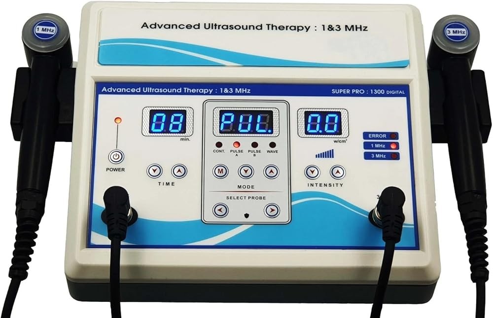
Nuwave Dual Frequency Therapeutic Ultrasound Device
Check Price on AmazonAs an Amazon Associate we earn from qualifying purchases.

SoundCare Plus Ultrasound Therapy Device
Check Price on AmazonAs an Amazon Associate we earn from qualifying purchases.
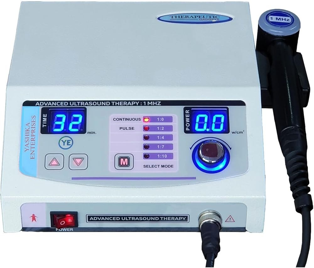
LGMedSupply Premium Ultrasound Device
Check Price on AmazonAs an Amazon Associate we earn from qualifying purchases.
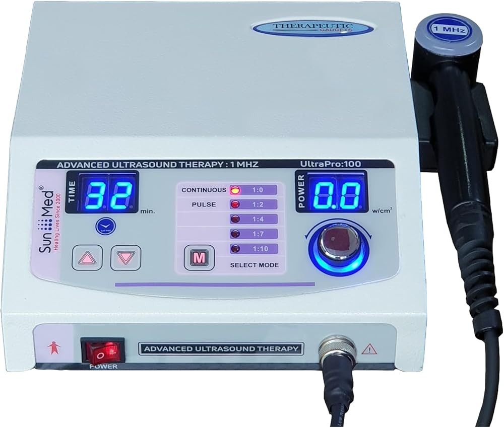
EverRelief Portable Ultrasound Therapy Machine
Check Price on AmazonAs an Amazon Associate we earn from qualifying purchases.
Recommended Products
Get Weekly Research Updates
New studies, updated reviews, and evidence-based health insights delivered to your inbox. Unsubscribe anytime.